The median umbilical ligament has. Obliterated umbilical artery median umbilical ligament.

Mcat Memoranda Umbilical Folds Median Medial And Lateral Are
The medial umbilical ligaments are anatomical remnants of the obliterated foetal umbilical arteries.
. The unobliterated medial umbilical ligament is defined by the presence of echogenic mucosal lines arrows along the course of both medial umbilical ligaments. The urachus connects the dome of the bladder to the umbilical cord during fetal life and is located behind the abdominal wall and anterior to the peritoneum in the space of Retzius. It is different from the median umbilical ligament a structure that represents the.
A fibrous cord extending from the urinary bladder to the navel that is the remnant of the fetal urachus. The key difference between medial and lateral is that medial is the term used to refer to structures close to the center or the median plane of an organism while lateral is the term used to refer to structures farther away from the median line. Median medial lateral.
Paired medial umbilical ligaments run along other side with a matching set of lateral ligaments. The urachus or median umbilical ligament represents the embryologic remnant of two embryologic structures. Paired medial umbilical ligaments run along other side with a matching set of lateral ligaments.
Access to the vesical pedicles and bladder was achieved through 2 windows on both sides. The medial umbilical ligament is the aforementioned paired structure related to the umbilical arteries while the median umbilical ligament contains the urachus. From lateral to medial.
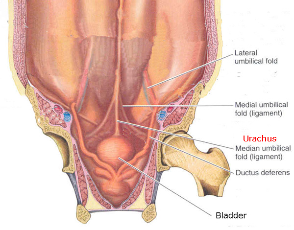
It then becomes the urachus in the fetus. The vertex of the bladder is joined to the umbilicus by the remains of the urachus which forms the middle umbilical ligament a fibromuscular cord broad at its attachment to the bladder but narrowing as it ascends. It extends from the apex of the bladder to the umbilicus on the deep surface of the anterior abdominal wall.
The median umbilical ligament begins as the allantois in the embryonic period. Lateral to this structure are the medial umbilical ligament and the lateral umbilical ligament. The median and medial umbilical ligaments form a peritoneal depression on each side of the urinary bladder referred to as the supravesical fossae.
Central intermediate while medial is of or pertaining to a mean or average. The image reveals a completely obliterated medial umbilical ligament without echogenic mucosal lines. Umbilical ligament medial a fibrous cord the remains of the obliterated umbilical artery running cranialward beside the bladder to the umbilicus.
It is important to distinguish between the medial vs median umbilical ligaments. Status of the medial umbilical ligament. The intercellular material or matrix is produced by the cells and gives the tissue its particular character.
Chronic abdominal pain is a very common condition that can have significant negative long-term psychosocial consequences including increased risk for anxiety school and work absences poor functional capacity and a. The supravesical fossa is the area of abdominal wall between remnant of urachus Median umbilical ligament and remnant of left or right umbilical artery medial umbilical ligament. The folds are 2 of the 5 umbilical folds and should not be confused with the single midline median umbilical fold.
It is on the deep surface of the anterior abdominal wall and is covered by the medial umbilical foldsplicae umbilicales mediales. Inferior epigastric vessels medial unbilical ligament. It is important to distinguish between the medial vs.
By birth the urachus is obliterated and becomes a vestigial structure known as the median umbilical ligament. The cloaca and the allantois. The remnants of an embryonic communication between the allantois and cloaca.
The median umbilical ligament is a structure in human anatomy. As adjectives the difference between median and medial is that median is situated in the middle. Inguinal swelling due to rare external supravesical hernia--a case report.
This later develops into the median umbilical ligament at birth. Lĭgəmənt strong band of white fibrous connective tissue connective tissue supportive tissue widely distributed in the body characterized by large amounts of intercellular substance and relatively few cells. The median arcuate ligament syndrome MALS is a cause of chronic abdominal pain affecting both children and adults alike.
The intraperitoneal view has the medial umbilical ligament as the lateral border of the bladder and the lateral umbilical ligament helps identify the inferior epigastric vessels. Medical Definition of median umbilical ligament. As nouns the difference between median and medial is that median is while medial is one or more letters that occur in the middle of a word.
Median umbilical ligament - Ligamentum umbilicale medianum. It is covered by the median umbilical fold. From superior to inferior.
Median is a related term of medial. The urachus extends from the anterosuperior surface of the bladder to the umbilicus and lies in the extraperitoneal space of Retzius or retropubic space between the transverse fascia. It is different to the median umbilical ligament a structure that.
The medial umbilical ligament is a paired structure found in human anatomy. Uteropelvic ls expansions of muscular tissue in the broad ligament of the uterus radiating from the fascia over the internal obturator muscle to the side of the uterus and the vagina. It is on the deep surface of the anterior abdominal wall and is covered by the medial umbilical folds.
It is also formed from the cloaca in utero. About Press Copyright Contact us Creators Advertise Developers Terms Privacy Policy Safety How YouTube works Test new features Press Copyright Contact us Creators. The medial umbilical ligaments are paired structures related to the umbilical arteries found either side of the median umbilical ligament.
Anterior muscles of leg The Hospitals. The median umbilical ligament runs down the lower portion of the front of the abdominal wall. The medial umbilical ligamentor cord of umbilical artery or obliterated umbilical artery is a paired structure found in human anatomy.
It is a shrivelled piece of tissue that represents the remnant of the embryonic urachus. Median plane or midline is the line drawn within the body in order to divide the body into right and left portions. The bilateral supravesical fossae lie between the median and bilateral medial umbilical folds.
The first between the. Vetebrae subtypes Certain Doctors Luv Saddling Coeds. Cervical Dorsal Lumbar Sacrum Coccyx.

Median Umbilical Ligament Wikipedia
![]()
Medial Umbilical Ligament Anatomy Branches Supply Kenhub

Medial Umbilical Ligament Wikipedia

Testpress Medical This Is One Of The Most Confusing Questions Asked In Embryology But For Those Who Can Spend Time To Understand It This Is For An Easy Retention And

Medial Umbilical Ligament Wikiwand
Epos Trade

Abdominal Wall Peritoneum And Intestines Lo 2 Abdominal Wall Youtube

Internal Abdominal Wall Inguinal Canal A Little Bit Of Thorax Flashcards Quizlet
0 comments
Post a Comment